These are some of the imaging and analytical techniques we use to reveal the methods and materials of ancient Egyptian craftsmen. Through these we discover the skill and ingenuity of the carpenters and artists, and the practical problems they faced.
The process of investigation begins with detailed examination, often using a stereomicroscope with magnification up to about ×60, and a raking light to show up surface relief.
Features of interest are recorded photographically using ultraviolet light to help locate organic coatings such as varnishes and clarify areas of restoration, and infrared radiation to reveal carbon-based pigments below the surface (e.g., in underdrawings). Visible-light induced luminescence (VIL) is a photographic technique that captures the infrared fluorescence of the pigment Egyptian blue.
Fibre optic reflectance spectroscopy (FORS) and X-ray fluorescence spectrometry (XRF) allow identification of pigments without the need to remove samples.
Further information can be obtained by taking tiny samples and analysing them with polarised light microscopy (PLM), X-ray powder diffraction (XRD) and Raman spectroscopy. The layer structure of the decoration is visible on cross-section samples viewed under high magnification. Analyzing these samples in a scanning electron microscope with energy-dispersive X-ray spectroscopy (SEM-EDX) locates the various inorganic pigments.
Original varnishes and resins on the coffins are identified from tiny samples using Fourier transform infrared spectroscopy (FTIR), which reveals the general character of the material, allowing us, for example, to differentiate tree resins from sugary gums. This is followed by gas chromatography–mass spectrometry (GC-MS), which can identify all the individual molecular components of the material so that its botanical origin can be determined.
X-radiography and Computed tomography (CT) scanning look beneath the surfaces of coffins to reveal the methods of construction, and scanning electron microscopy is used to identify all the different woods employed.
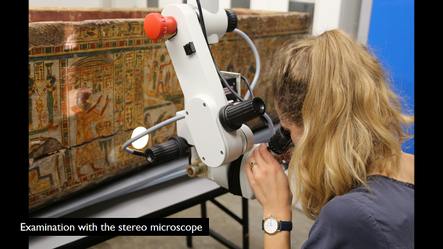
A conservator examines the outer coffin box of Nespawershefyt with a stereomicroscope. Fitzwilliam Museum collection: E.1.1822.
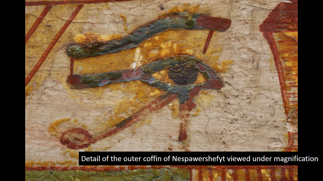
Detail of a wedjat-eye from Nespawershefyt's outer coffin box viewed under magnification.
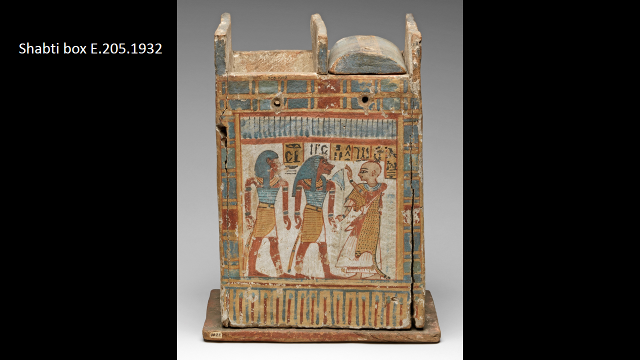
New Kingdom shabti box from the Fitzwilliam Museum collection: E.205.1932.
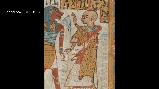
Detail of the shabti box owner.
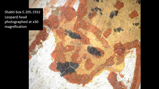
Detail of the leopard head worn by the shabti box owner at x30 magnification.
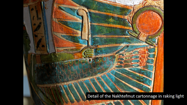
Detail of the cartonnage of Nakhtefmut under raking light. Fitzwilliam Museum collection: E.64.1896.
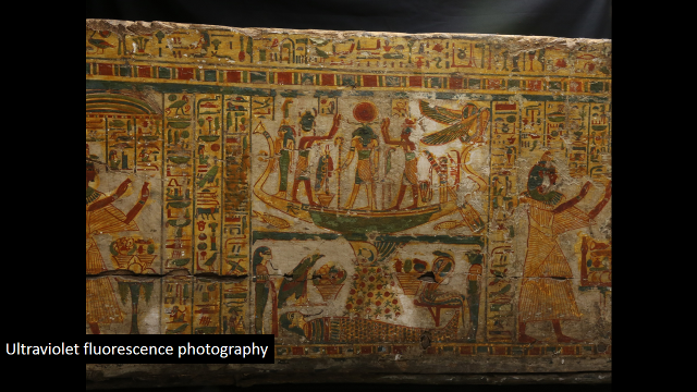
Detail of the outer coffin box of Nespawershefyt. Fitzwilliam Museum collection: E.1.1822.
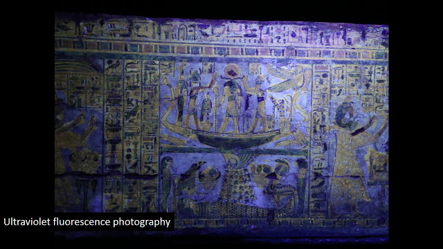
Nespawershefyt's outer coffin box under ultraviolet fluorescence light.
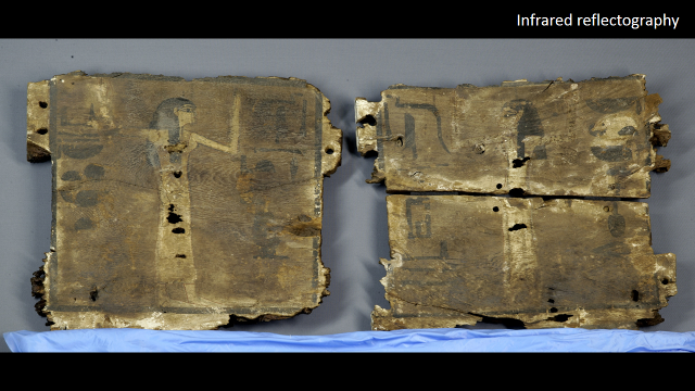
Coffin fragments of the mourning women. Fitzwilliam Museum collection: E.283.1900.
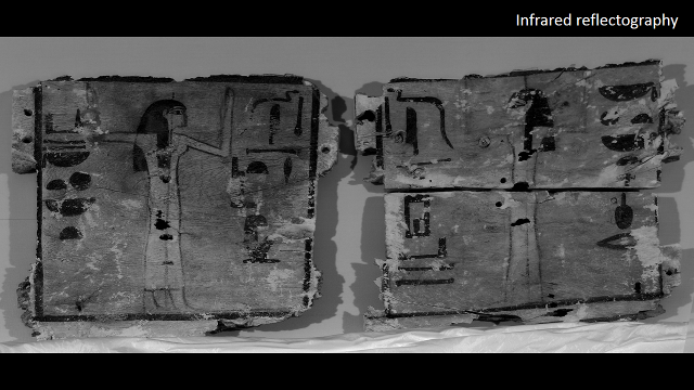
Infrared reflectography applied to the coffin fragments of the mourning women.
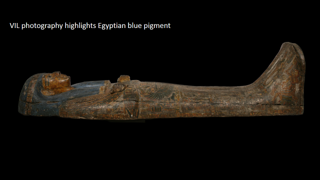
Inner coffin lid of Nespawershefyt. Fitzwilliam Museum collection: E.1.1822.
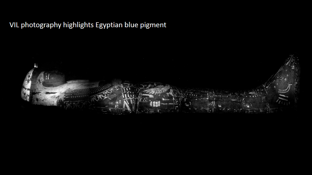
Application of VIL photography highlighting Egyptian blue pigment on the inner coffin lid of Nespawershefyt.
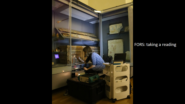
Conservator Jenny Marchant examining a Middle Kingdom box coffin using FORS.
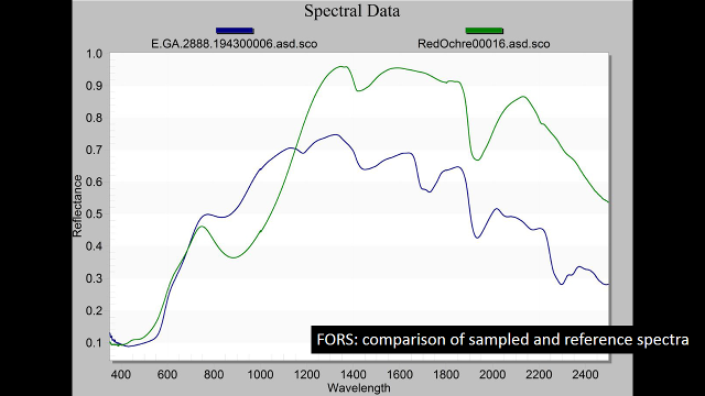
A FORS reading showing a comparison of sampled and reference spectra.
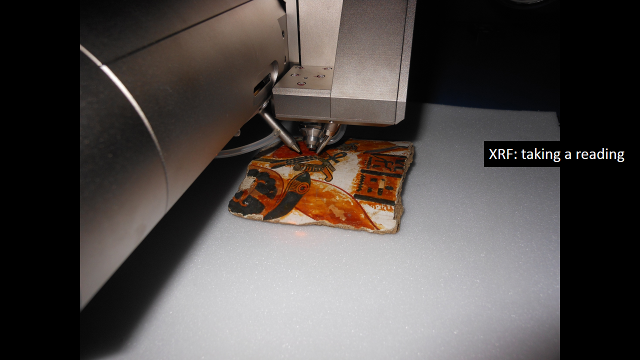
Taking a reading using XRF.
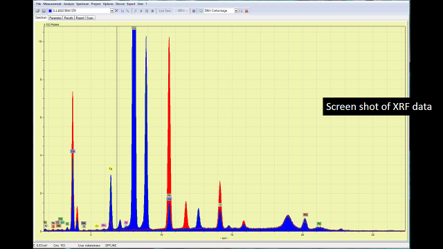
Screenshot showing the output of XRF data.
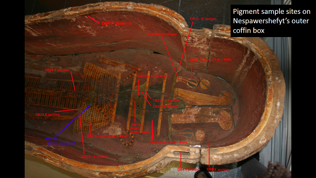
Pigment sample sites on Nespawershefyt's outer coffin box. Fitzwilliam Museum collection: E.1.1822.
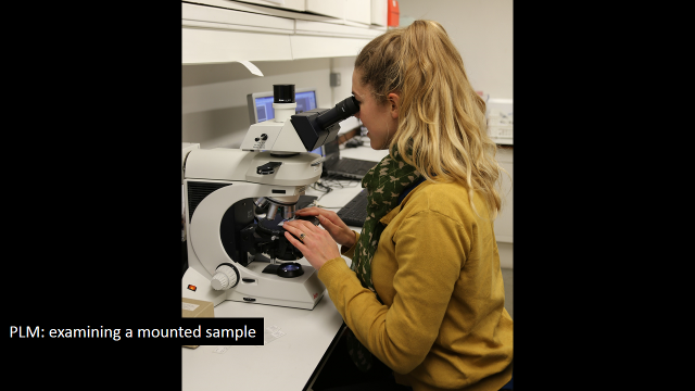
A conservator examines a mounted sample using PLM (Polarised Light Microscopy).
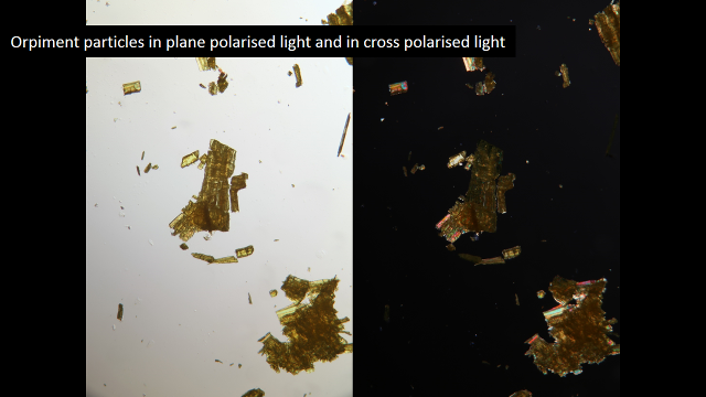
Particles of orpiment pigment in plane polarised light and cross polarised light.
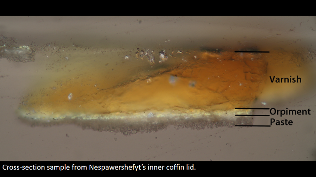
Cross-section sample from Nespawershefyt's inner coffin lid.
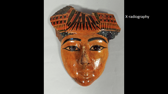
Fragment of a face from a yellow coffin. Fitzwilliam Museum collection: E.GA.507.1947.
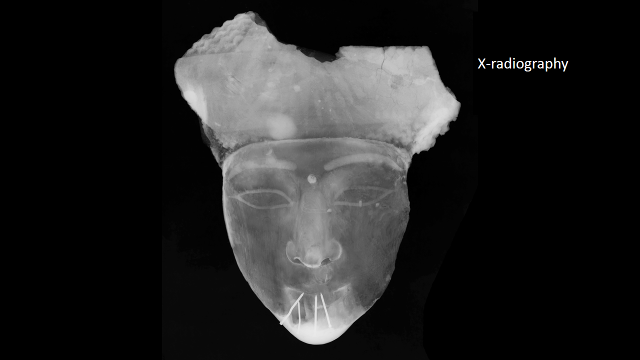
X-radiograph of a coffin fragment showing modern repair work in the form of metal nails in the chin.
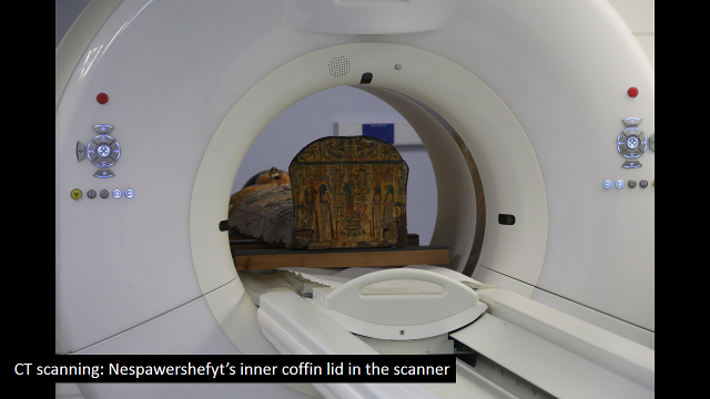
CT-scanning the inner coffin lid of Nespawershefyt at Addenbrookes Hospital.
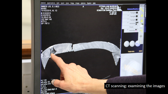
Examining the images of the CT-scans.
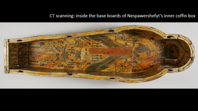
Inside the inner coffin box of Nespawershefyt. Fitzwilliam Museum collection: E.1.1822.
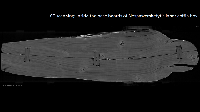
CT-scan of the base of Nespawershefyt's inner coffin box showing mortise and tenon joints.
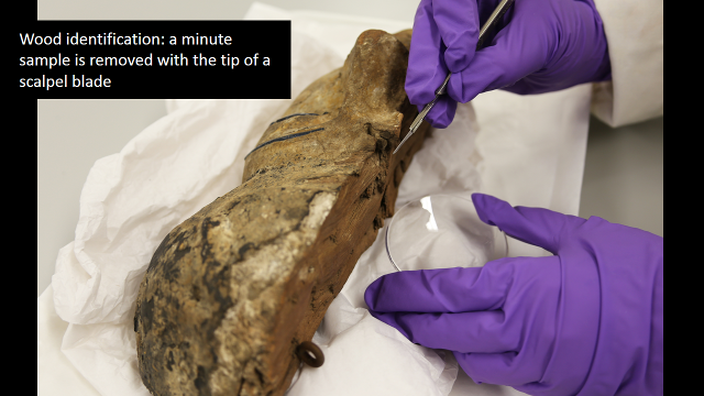
Wood sampling using a scalpal blade.
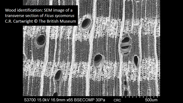
SEM image of a transverse section of *Ficus sycomorus* wood. Copyright Caroline Cartwright: British Museum.